Researchers discover: This is how microtubuli can organize themselves in growing nerve cells
For nerve fibres (neurons) to be able to transport proteins and other crucial materials along their conducting processes (axons), a directional carrier system is required. This system consists of rod-like molecules, so-called microtubules. Researchers at the University of Cambridge and the Max Planck Zentrum für Physik und Medizin have now been able to demonstrate, for the first time, how microtubules are specifically aligned within growing axons by combining molecular biological methods with physical modelling and computer simulations.
For nerve fibres (neurons) to be able to transport proteins and other crucial materials along their conducting processes (axons), a directional carrier system is required. This system consists of rod-like molecules, so-called microtubules. Researchers at the University of Cambridge and the Max-Planck-Zentrum für Physik und Medizin have now been able to demonstrate, for the first time, how microtubules are specifically aligned within growing axons by combining molecular biological methods with physical modelling and computer simulations.
For humans to be able to wiggle their toes, messages need to travel from the brain to the foot, a distance well over a meter in many adults. This is made possible by neurons, which transmit electrical signals along long extensions called “axons”. Axons can only function properly if all the required molecules produced in a part of the neuron known as the cell body, are ferried to their opposite ends. This ferrying around of molecules is done along long, filamentous molecules called microtubules, which act as a directed highway along which molecules are shuttled, either towards or away from the cell body.
Microtubules can be thought of as asymmetrical rods. One end – known as the plus end – is dynamic and can undergo growth or shrinkage, while the other end – called the minus end – is stable. For transport along the axon to happen efficiently, microtubules in the neuron need to be oriented with their plus end pointing towards the ends of the axon. Microtubules in growing neurons develop this orientation, but how that is achieved is not fully understood.
To understand the basis of this cellular phenomenon, Maximilian Jakobs (University of Cambridge), Assaf Zemel (Hebrew University of Jerusalem) and Kristian Franze (University of Cambridge, FAU Erlangen-Nürnberg & Max Planck Zentrum für Physik und Medizin) examined the behaviour of microtubules in developing neurons from fruit fly larvae. A fluorescent protein, which emits light when the microtubules are growing, helped the researchers visualise the growing plus end of microtubules and microtubule orientations in developing axons. These experiments showed that microtubules whose plus end pointed towards the axon end shrank less frequently than those with opposite orientation, leading to their enhanced growth. This resulted in a higher proportion of the correctly-oriented microtubules in the axon.
Treating the neurons with Nocodazole, a chemical that disrupts microtubule growth, or with sodium chloride, which changes the osmotic pressure, decreased axon growth and disrupted the uniform orientation of the microtubules in the axon, unravelling a link between the growth of microtubules and their uniform orientation in axons.
The next step was to determine whether specific axonal proteins such as p150 – a protein that is enriched at the tip of the axon and decreases microtubule shrinkage rates – are involved in this process. Reducing the levels of p150 in fruit flies using molecular and genetic methods resulted in microtubules with their plus end pointing towards the axon tip shrinking more frequently, reducing their lengths and thus the proportion of microtubules with this orientation in the axon. This role of proteins enriched in the axonal tip, along with previously discovered mechanisms, explains how microtubules align unidirectionally in axons.
It was shown that proteins, which are enriched at the axon tip and which regulate microtubule growth, such as the protein p150, are important for the uniform orientation of microtubules in neuronal axons. Perturbations of microtubule growth leads to a decrease in axonal microtubule orientation, which is critical for the function of healthy neurons.
These findings open new avenues of research into neurodegenerative diseases like Alzheimer’s and Parkinson’s, which might manifest due to a breakdown of transport along microtubules in neurons.
Original publication: Maximilian AH Jakobs Assaf Zemel Kristian Franze (2022) Unrestrained growth of correctly oriented microtubules instructs axonal microtubule orientation eLife 11:e77608.
https://doi.org/10.7554/eLife.77608
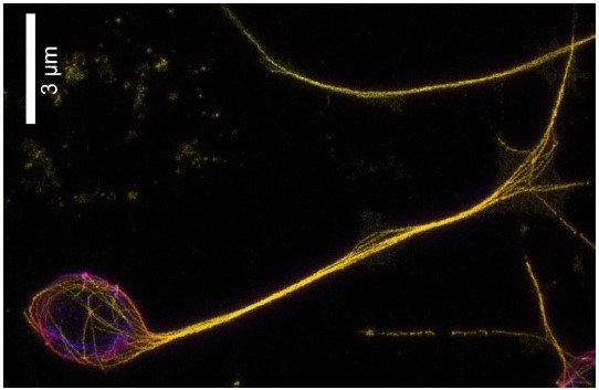
Picture (Reprinted with permission from Maximilian AH Jakobs et al., elifesciences.org 2022): Expansion microscopy image of D. melanogaster neuron; microtubules are stained in yellow.
Contact
Edda Fischer
Head of Communication and Marketing
Telefon: 09131 7133 805
MPLpresse@mpl.mpg.de